Revolutionizing Dental Care with 3D Imaging: An In-Depth Look at Cone Beam Scanners featuring Kristen Campbell DDS
The dental field is constantly innovating and improving, and chances are, your latest visit to the dentist will look quite a bit different from visits you made five or ten years ago. Cone beam scanners–also referred to as cone beam CT or CBCT–is one such advancement making dental services and treatment more convenient, more comfortable, and more precise. This imaging technology replaces traditional CT scans and some types of radiography to more accurately identify problem areas in the mouth and assist dentists in creating effective treatment plans.
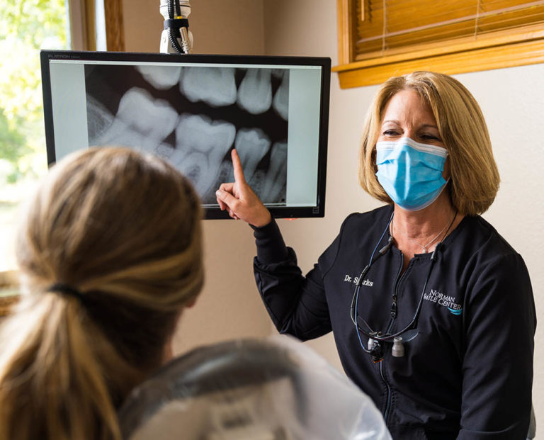
Dr. Kristin Campbell and Dr. Donna Sparks of Norman Smile Center have spent their careers learning and mastering the latest techniques, technology, and procedures in dentistry so they can provide their patients with the best experience possible. They’ve been serving pediatric, teen, and adult patients in Norman and the surrounding area for more than 30 years and offer the latest in precision dentistry, diagnostics, and digital imaging like cone beam scanners.
Dental Cone Beam Scanners: The Basics
A dental cone beam CT is sometimes called dental cone beam computed tomography (CBCT). Like other types of CT scans, a dental cone beam will allow dentists to see the interior structure of your face so that they can identify potential problems that they can’t see from the outside.

How Dental CBCT Works
A dental cone beam CT sends an invisible beam into the areas at which it is pointed. These beams travel through or bounce off of different materials in the body, and the CT scanner interprets this information to determine what those structures look like. A cone beam CT, as the name suggests, sends those beams out in a rotating cone shape, then calculates everything that has been exposed to the scan based on how the beams behaved and produces a detailed 3D computer graphic.
This is in contrast to a traditional CT, which sends out beams in a fan-like shape so its images are created like thin “slices” that are then stacked together to create an image, resulting in less detail, both in terms of depth and also total image capacity.
The Dental Cone Beam Scan Process
Getting a dental cone beam CT is virtually painless, requiring no contact with the machine in order to obtain high-quality, highly-detailed images. You will rest your head on a specially designed mount that is comfortable for your chin, helping ensure that your head stays in the right position and doesn’t move while images are being captured. (Movement during a CBCT scan can make the images inaccurate or difficult to interpret.) You might be asked to bite down on a thin piece of plastic to prevent your mouth from shifting while pictures are being taken. Then, a round or rectangular beam projector will rotate 360 degrees around your head, gathering images from all angles of your mouth for a more accurate picture. A cone beam CT can be completed in your dentist’s office in less than a minute and produces no lights, sounds, or physical sensations.
Use Cases for Dental Cone Beam Scanners
Dental cone beam CT scans help dentists like Dr. Campbell and Dr. Sparks of Norman Smile Center improve the type and quality of patient care and treatment. These types of scans can more accurately depict the parts of the mouth, face, and jaw that a dentist can’t see, like your jawbone, tooth roots, and nerves, allowing for more precision in diagnoses and more thorough assessments.
Improved Precision for Dental Implants
Dental cone beam CTs are ideal for detailed and invasive procedures like dental implants. Implants are placed by inserting a tiny titanium post directly into the jawbone, where the metal then fuses with the bone in a process called osseointegration. Then, once the post is stable, the artificial tooth (or crown) is attached to it and functions just like a normal tooth.
Older imaging methods lack the level of detail of cone beam CTs, increasing the chances that an implant is misplaced or placed in a part of the jaw with inadequate density. The 3D image produced by cone beam CTs help make the dental implant procedure much more reliable and predictable, allowing the dentist to see everything from whether your jawbone is thick enough for an implant and the ideal place to insert it to whether the bone has properly fused with the abutment, all from a computer screen.
More Accurate Cosmetic Procedures
Cone beam CT scans are also useful for cosmetic procedures. The goals of a cosmetic procedure are to improve the smile and the visual appearance of the face, both of which requires dentists to be able to predict what will happen when they make changes. Cone beam CTs can provide a 3D image that dentists can manipulate or modify in order to accurately reflect how treatments, additions, and alterations will look once the procedure is complete. It also gives them as much information as possible on what’s already there so that they know exactly how to achieve the results that you want.
Detailed Diagnostics
The most common use of dental cone beam CTs is for diagnostic purposes. Being able to see the structure of your jawbone and even some of the areas that are more difficult to image—like nerves and sinuses—helps dentists to spot problems long before they develop into something significant. This is especially true for issues that are primarily uncovered using 3D imaging, like bone infections and temporomandibular joint disorders.
Revolutionizing Patient Care with Dental Cone Beam Scanners
Cone beam CTs have a number of advantages over traditional CT scans, including:
- Image quality. One of the primary advantages of cone beam CTs is the quality of the images they produce. While traditional CT scans can only generate still images, cone beam scanners produce 3D computer graphics that can be moved around, rotated, and manipulated right on the screen, providing a better understanding of things like how thick your jawbone is or how much space your nerves are occupying.
- Reduced radiation exposure. Whenever you have imaging done, you’ll be exposed to some level of radiation. That’s why you’re asked to wear those heavy vests when you get an x-ray—the technician is attempting to reduce the radiation that you are exposed to as much as possible. Cone beam scanners produce almost no radiation, exposing you and your technicians to ten times less radiation on average than traditional CT machines.
- Virtual modeling. Dentists can use the 3D computer-generated graphics produced by cone beam scanners to help patients visualize how dental procedures and cosmetic options will impact their smiles. They can also make changes to the virtual model to demonstrate in real time how changes will look.
Norman Smile Center: Your Trusted Provider for the Latest in Dental Care
Dr. Campbell and Dr. Sparks, along with their team at Norman Smile Center, are proud to be at the forefront of dental technology so we can deliver comprehensive preventative, cosmetic, and restorative dental services with consistent, reliable results. Whether you’re considering a cosmetic procedure or need help keeping your oral health in good shape, Norman Smile Center has the imaging tools and technical experience you need for accurate diagnoses and effective treatments that will keep your smile healthy and beautiful for a lifetime. Find out how advanced technology like cone beam scanners can benefit you and schedule an appointment today!